What is Perthes Disease?
Legg-Calvé-Perthes disease is a condition in which blood supply to the femoral head is disrupted, leading to avascular (non-infectious) necrosis. It most commonly affects children 3-12 years old (peak incidence 5-7 years), predominantly boys (4:1 ratio).
The disease progresses cyclically: necrosis → fragmentation → healing → remodeling. The natural course takes 2-5 years. The primary concern is deformity of the femoral head, which can lead to early osteoarthritis in adulthood.
Stages of disease (Waldenström classification):
- I — Necrosis — disruption of blood supply, beginning of destruction. X-rays may appear normal
- II — Fragmentation — femoral head breaks down, areas of lysis appear. The most critical period
- III — Healing — new bone tissue growth, reossification
- IV — Remodeling — final shaping of the head. Deformity (coxa plana, coxa magna) determines long-term prognosis
Facts about Perthes Disease
- ICD-10: M91.1
- Age: 3-12 years (peak 5-7)
- Gender: boys 4 times more often
- Bilateral: 10-15% of cases
- Duration: 2-5 years
- Risk: early hip osteoarthritis with deformity
Symptoms of Perthes Disease
Early signs
- Limping (child limps without apparent cause)
- Pain in the groin, thigh, or knee
- Limited hip rotation and abduction
- Fatigue with walking
- Muscle spasm
Progression
- Limb shortening
- Atrophy of thigh and buttock muscles
- Marked limitation of movement
- Inability to run and jump
- Positive Trendelenburg sign
Prognostic factors
- Age of onset — before age 6 has better prognosis
- Degree of involvement — Herring classification (A, B, C)
- Preservation of shape — the less deformation, the better
- Early treatment — the key to a good outcome
How MIBRAR® helps with Perthes Disease
MIBRAR® accelerates natural bone regeneration, improves blood supply to the necrosis area, and helps prevent deformation of the femoral head — the main long-term problem of Perthes Disease.
Stage Assessment
MRI of the hip joint is analyzed to determine the disease stage, extent of involvement (Herring classification), and prognosis. An individual treatment plan is developed.
Obtaining Concentrates
From blood — CGF with angiogenic factors (VEGF, PDGF) for restoration of blood supply. From adipose tissue — Lipogems® with stem cells to accelerate osteogenesis.
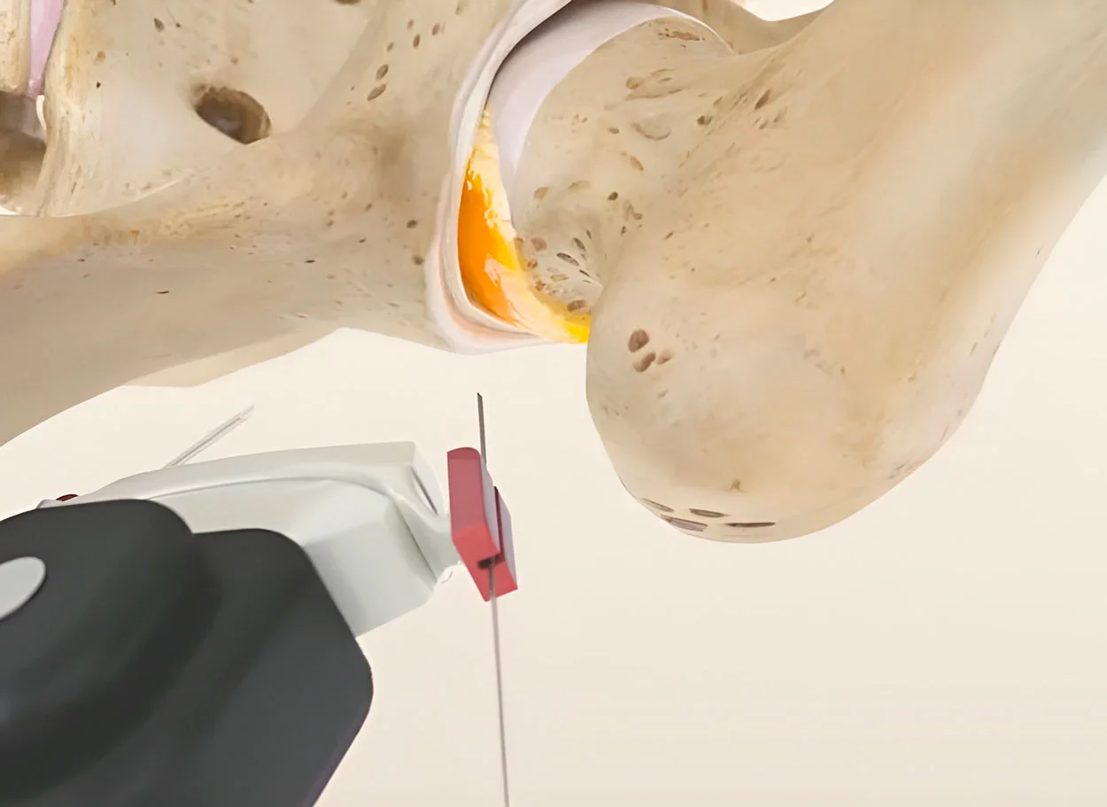
Injection into Necrosis Area
Under navigational control, concentrates are delivered directly into the femoral head — the area of disrupted blood supply. The procedure is minimally invasive and safe.
Accelerated Recovery
Stem cells and growth factors stimulate neovascularization and osteogenesis. This accelerates the recovery phase and promotes formation of a more spherical femoral head, reducing the risk of deformation and early osteoarthritis.
Scientific Basis of MIBRAR® for Perthes Disease
Traditional treatment of Perthes Disease involves observation, orthoses, activity restriction, and in severe cases — surgery (osteotomy). None of these methods accelerate bone regeneration. MIBRAR® fills this gap — delivering stem cells and growth factors directly to the necrosis area.
How MIBRAR® Affects Perthes Disease:
- Neovascularization — vascular growth factors (VEGF, PDGF, FGF) stimulate growth of new blood vessels in the ischemic area, restoring blood supply to the femoral head
- Osteogenesis — mesenchymal stem cells differentiate into osteoblasts and accelerate formation of new bone tissue in the recovery phase
- Anti-inflammatory effect — reduction of synovitis and edema typical of the active disease phase
- Preservation of shape — accelerated regeneration helps maintain a more spherical femoral head shape, reducing the risk of deformation (coxa plana)
Important: MIBRAR® is most effective in early stages and the fragmentation phase, when natural recovery begins. Each case is evaluated individually.
Advantages of MIBRAR® for Perthes Disease
- Accelerated recovery — shortened disease duration
- Deformation prevention — preservation of femoral head shape
- Minimal invasiveness — micro-puncture
- Without general anesthesia — local anesthesia
- Osteoarthritis protection — long-term prognosis
MIBRAR® Method Advantages
95% of interventions covered
MIBRAR® covers up to 95% of all spinal neurosurgery and orthopedic operations.
No anesthesia or incisions
Outpatient treatment via 0.3-1.5 mm puncture. No general anesthesia or hospitalization.
No age restrictions
Regeneration at any age. Safe for chronic conditions and anesthesia intolerance.
Rapid improvement
Concentrates have analgesic and anti-inflammatory properties. Relief within days.
Multiple zones at once
Simultaneous treatment of multiple discs or joints in one procedure.
Home the same day
No crutches, braces or rehabilitation needed. MRI follow-up at 8-16 weeks.
MIBRAR® Technology
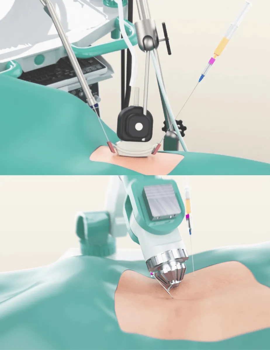
Cyber Navi Hand™
Intraoperative robotic navigation system. Provides precise access to deep structures with 1 mm and 1 degree accuracy.

Sono Control Arm™
Device for intervention under sonographic control. Eliminates open surgeries with real-time visual monitoring.