What is a meniscus tear?
The menisci are C-shaped cartilage pads located between the femur and tibia in the knee joint. Each knee has two menisci — the medial (inner) and lateral (outer) meniscus. They function to absorb shock, stabilize the joint, and distribute load.
Meniscus tear is one of the most common knee injuries. Over 400,000 knee arthroscopies are performed annually in Germany, with the majority for meniscal issues. However, scientific research has shown that meniscectomy accelerates osteoarthritis development 3-6 times faster.
Types of tears:
- Longitudinal (vertical) — along the fiber direction
- Horizontal — meniscal delamination
- Radial — transverse tear
- Bucket-handle — flap tear
- Degenerative — age-related wear without clear trauma
- Complex — combination of multiple tear types
Vascular zones:
- Red zone (periphery) — good blood supply, capable of healing
- Red-white zone — limited blood supply
- White zone (inner) — virtually no blood vessels, does not heal spontaneously
Meniscus Facts
- ICD-10: M23.3 / S83.2
- Incidence: 60-70 cases per 100,000 per year
- Medial: injured 3 times more often
- Arthroscopies: 400,000+/year in Germany
- After meniscectomy: osteoarthritis 3-6 times faster
Symptoms of meniscus tear
Acute tear (traumatic)
- Sharp pain at the moment of injury
- Clicking or popping sensation in the knee
- Rapid swelling (within hours)
- Joint locking (inability to fully extend)
- Inability to bear full weight
Degenerative tear
- Gradually increasing pain
- Pain along the joint line
- Intermittent locking
- Swelling after activity
- Crunching and clicking with movement
- Night pain
Characteristic Tests
- Pain with squatting
- Pain with knee twisting
- Tenderness on palpation of the joint line
- Positive McMurray test
- Quadriceps muscle atrophy
Arthroscopy vs MIBRAR®
| Criterion | Arthroscopic Resection | Arthroscopic Repair | MIBRAR® |
|---|---|---|---|
| Principle | Removal of damaged portion | Suturing the tear | Meniscus tissue regeneration with stem cells |
| General anesthesia | General / spinal | General / spinal | Without anesthesia |
| Meniscus Preservation | ❌ Meniscus reduced | ✅ If healing occurs | ✅ Fully preserved + regeneration |
| Osteoarthritis Risk | Increased 3-6 times | Lower than resection | Reduced — meniscus restored |
| White Zone | Resection only | Suture often fails to heal | ✅ Stem cells create blood supply |
| Return to Sports | 4-6 weeks | 3-6 months | 4-8 weeks |
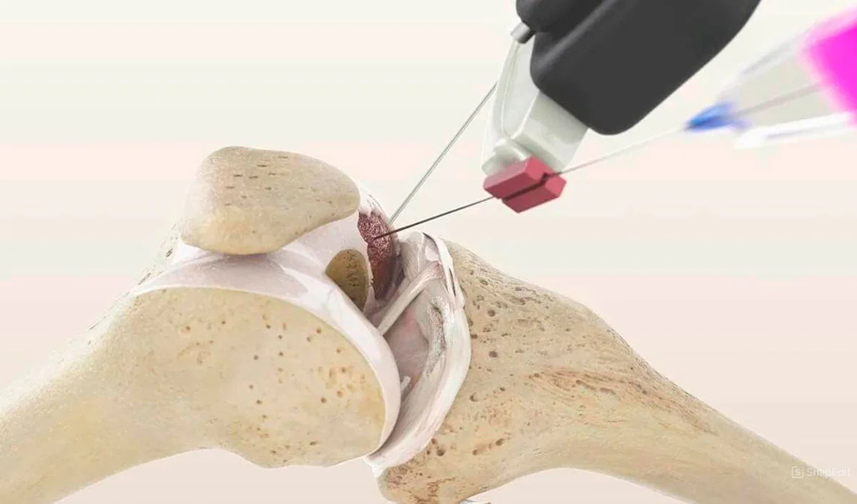
How MIBRAR® regenerates the meniscus
MRI Diagnosis
MRI of the Knee Joint is analyzed using Cyber Navi Hand™. Tear type, location (red/white zone), and associated articular cartilage and ligament damage are determined.
Biomaterial Harvesting
CGF from blood — growth factors to stimulate healing. Lipogems® from adipose tissue — stem cells capable of differentiating into meniscal chondrocytes.
Targeted Injection
Under Sono Control Arm™ guidance, autologous concentrates are injected directly into the meniscus tear zone. Key advantage: stem cells create neovascularization in the white zone, which typically does not heal.
Regeneration
Stem cells form new meniscus tissue in the tear zone. Growth factors stimulate angiogenesis and collagenogenesis. After 3-6 months — meniscus integrity restoration, visible on follow-up MRI.
Why MIBRAR® regenerates the meniscus
The main problem with the meniscus is the "white zone" (inner 2/3), which is practically devoid of blood vessels and incapable of spontaneous healing. This is why surgeons remove the damaged portion. But MIBRAR® solves this problem biologically — stem cells create new vessels in the avascular zone.
Meniscus Regeneration Mechanism:
- Neovascularization of the white zone — vascular growth factors (VEGF) from CGF stimulate capillary growth in the avascular meniscus portion, creating healing conditions
- Chondrogenesis — stem cells from Lipogems® differentiate into meniscal chondrocytes and synthesize type I and II collagen
- Scar Formation — fibroblasts fill the tear zone, forming strong tissue
- Anti-inflammatory Effect — suppression of inflammation caused by free meniscus fragments
Result: meniscus preserved and restored. This is critically important, as removal of even 20% of the meniscus increases contact pressure on the cartilage by 350%, inevitably leading to osteoarthritis.
Why It’s Important to Preserve the Meniscus
- 70% of the load on the knee goes through the menisci
- Resection of 20% → pressure +350%
- Osteoarthritis 3-6 times faster after resection
- MIBRAR®: preservation + regeneration
Treatment Results for Meniscus Tear
Follow-up MRI scans confirm complete meniscus regeneration after the MIBRAR® procedure — without resection or arthroscopy.

MIBRAR Case: Male, 56 years
Diagnosis: Complex posterior horn medial meniscus tear, patellofemoral and tibiofemoral cartilage wear, grade I-II arthrosis. 2 years of pain during walking and bending, swelling, night pain.
Result at 4 weeks: MRI shows healed meniscal tissue (green oval). One week post-surgery — no complaints. After 3 weeks — sports without restrictions.
View case study →
Same patient — sagittal view
Sagittal MRI: before treatment — tear line in the posterior horn of the meniscus (circled in green). Tibiofemoral cartilage degeneration.
At 4 weeks: New cartilage layer formed through reconstruction. Meniscus fully restored through regeneration — resection was not needed.
View case study →
Same Patient — Third Sagittal Slice
Sagittal MRI (image 5): before treatment — tear zone in the posterior horn of the meniscus (green oval). Femoral condyle cartilage surface also damaged.
After 4 weeks (image 6): meniscus integrity restored, tear zone filled with new tissue. Three MRI slices of one patient confirm: MIBRAR® provides complete meniscus regeneration, not scarring.
View case study →MIBRAR® Method Advantages
95% of interventions covered
MIBRAR® covers up to 95% of all spinal neurosurgery and orthopedic operations.
No anesthesia or incisions
Outpatient treatment via 0.3-1.5 mm puncture. No general anesthesia or hospitalization.
No age restrictions
Regeneration at any age. Safe for chronic conditions and anesthesia intolerance.
Rapid improvement
Concentrates have analgesic and anti-inflammatory properties. Relief within days.
Multiple zones at once
Simultaneous treatment of multiple discs or joints in one procedure.
Home the same day
No crutches, braces or rehabilitation needed. MRI follow-up at 8-16 weeks.
MIBRAR® Technology
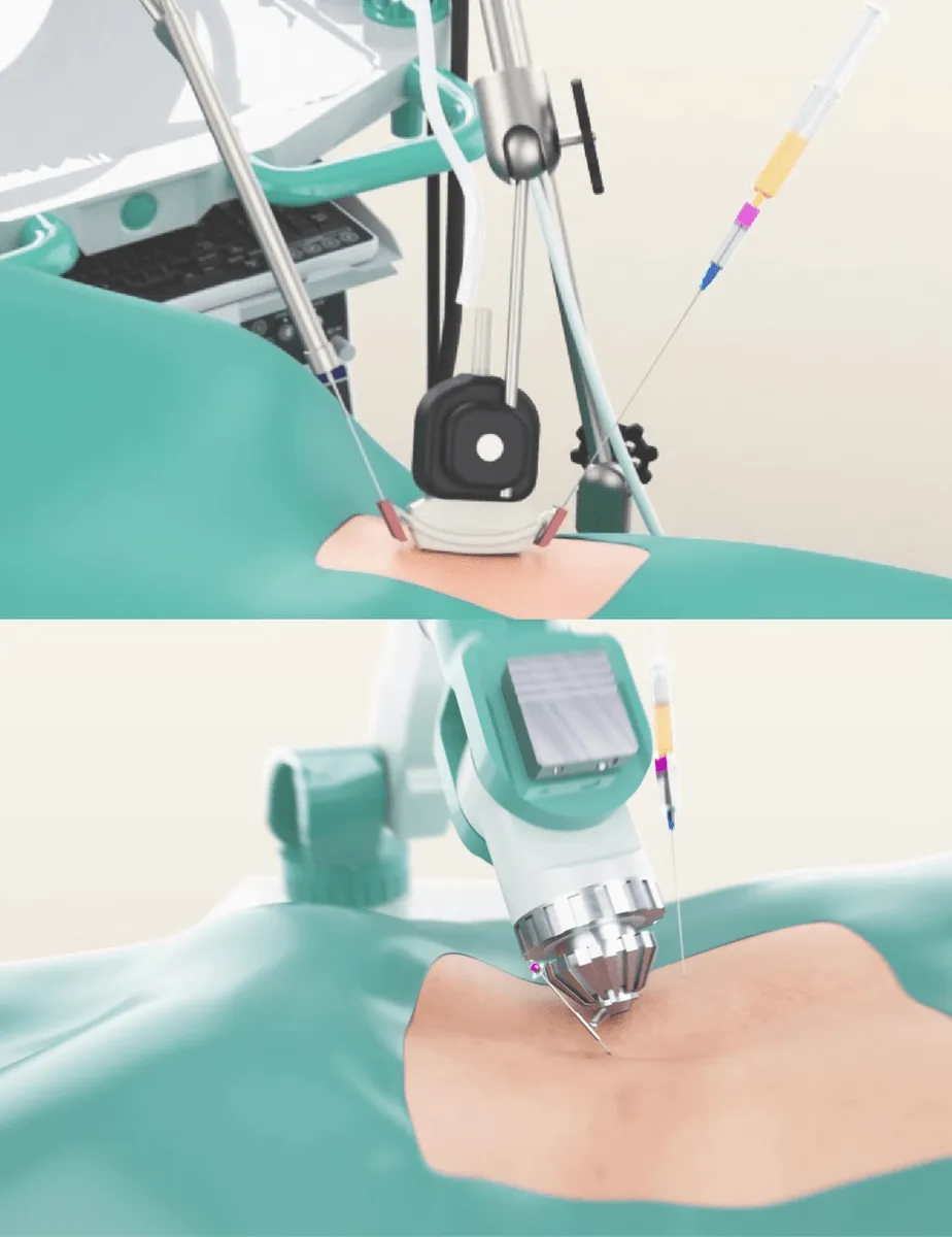
Cyber Navi Hand™
Intraoperative robotic navigation system. Provides precise access to deep structures with 1 mm and 1 degree accuracy.

Sono Control Arm™
Device for intervention under sonographic control. Eliminates open surgeries with real-time visual monitoring.
