What intradiscal regenerative therapy is
It is the targeted injection of autologous biopreparations (CGF, PRP, Lipogems®, BMAC) into spinal structures — intervertebral disc, epidural space, facet joints, sacroiliac joint — under fluoroscopic guidance or CT navigation. The aim is pathogenetic treatment of degenerative spinal disease without surgery. Unlike classical epidural steroid blocks (which only mask symptoms), regenerative therapy restores disc structure, relieves neuroinflammation and stimulates tissue remodelling. The patient returns to normal life the day after the procedure.
Indications for intradiscal therapy
The spectrum of indications is broad — from early disc degeneration to severe FBSS. The key principle is accurate identification of the pain generator via MRI, provocative discography or diagnostic blocks. The choice of technique depends on this: intradiscal, epidural, facet or intraosseous.
Degenerative disc disease (DDD)
Pfirrmann II–IV — disc dehydration, height loss, discogenic pain. Intradiscal CGF/PRP/Lipogems® restores disc hydration up to 25% and reduces pain by 60–80%.
Disc herniation up to 15 mm without sequestration
Protrusions and extrusions at L4–L5, L5–S1, C5–C6, C6–C7. Epidural PRP + intradiscal CGF — 75–85% avoid surgery and achieve significant improvement within 3–6 months.
Radicular syndrome (radiculopathy)
Pain radiating to leg/arm without progressive paresis. Transforaminal epidural CGF injection — an alternative to steroid block without side effects.
Facet syndrome (spondyloarthrosis)
Arthrosis of the facet joints. Local pain that worsens on extension. Regenerative facet blocks with CGF/Lipogems® under fluoroscopic guidance.
FBSS (Failed Back Surgery Syndrome)
Persisting or recurrent pain after laminectomy, discectomy, fusion. Epidural neuroplasty + Lipogems® into the scar–adhesive fibrosis area. Effectiveness 65–75%.
SI joint dysfunction (sacroiliitis)
Local pain in the sacroiliac joint, radiating to the buttock. A frequently missed cause of "lumbar pain". Fluoroscopy-guided CGF — a precise and effective alternative to a steroid block.
Modic II–III disease
Endplate changes on vertebral bodies on MRI — a frequent cause of chronic discogenic pain. Intraosseous BMAC injection into the subchondral zone of Modic changes reduces pain in 75% of cases.
Injection techniques
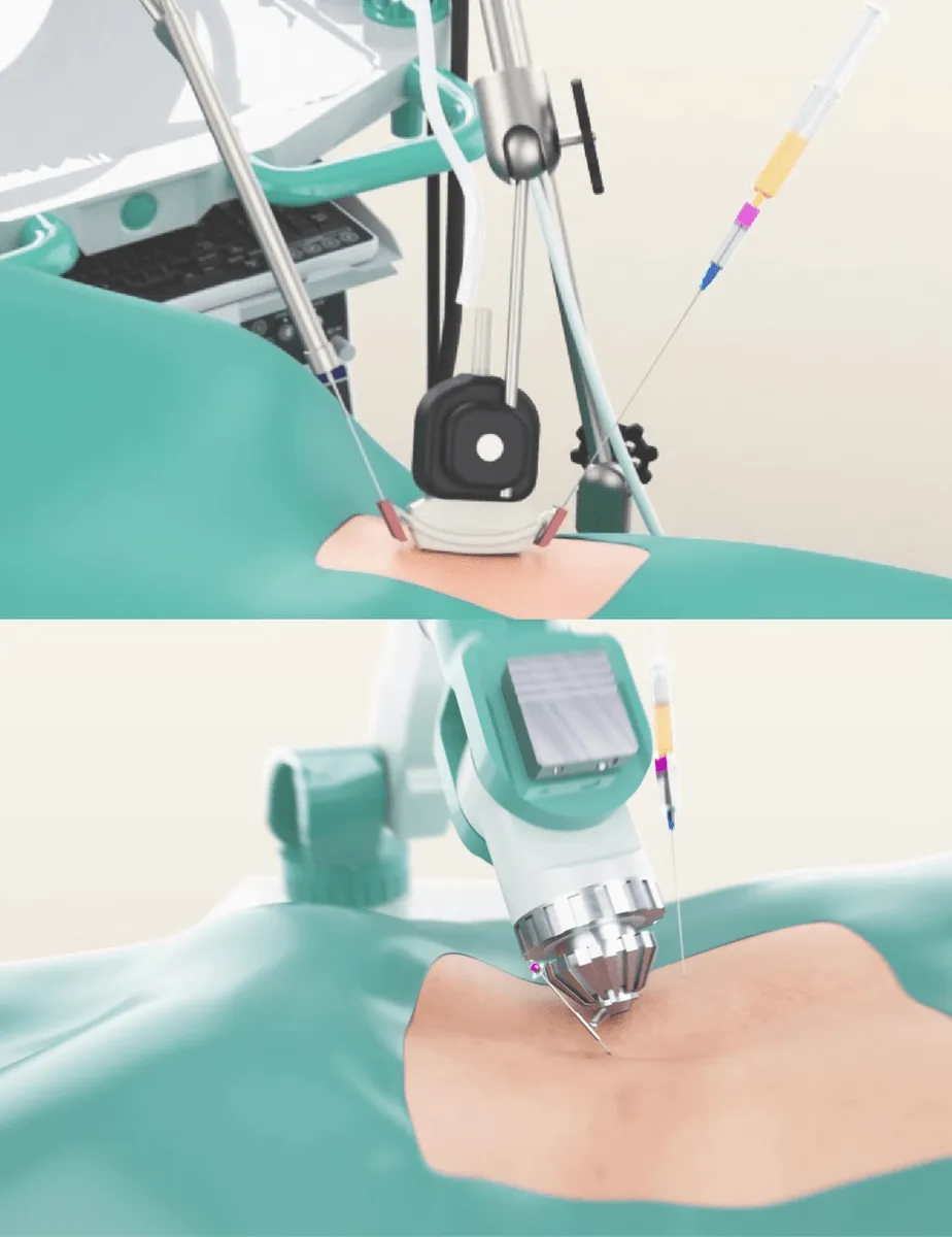
Intradiscal
Injection into the nucleus pulposus through a thin (22G) needle under fluoroscopic guidance. Postero-lateral access through Kambin\u2019s triangle (for lumbar levels), a safe path without root injury. Duration 15–20 minutes per level.
Transforaminal epidural
Injection into the epidural space through the foraminal window under fluoroscopy with contrast verification. Precise delivery of CGF to the inflamed root. An alternative to steroid block without side effects.
Caudal epidural
Injection through the sacral hiatus — the safest epidural injection technique. Used in multilevel lumbar problems. Neuroplasty (adhesiolysis) is possible in FBSS.
Facet block
Injection of CGF/Lipogems® into the facet joint cavity under fluoroscopic guidance. Used for spondyloarthrosis. Often combined with radiofrequency denervation of the medial branch to prolong the effect.
Sacroiliac injection
Injection into the sacroiliac joint through the lower third — the narrowest part. Under fluoroscopic guidance with contrast. For SI joint dysfunction, sacroiliitis, after spinal fusion.
Intraosseous
BMAC injection into the subchondral bone of endplates in Modic II–III disease, into compression fracture zones in osteoporosis, into avascular necrosis areas. The most innovative technique of the last decade.
Comparison with other methods
| Parameter | Steroid block | Microdiscectomy | Intradiscal regenerative |
|---|---|---|---|
| Effect duration | 4–12 weeks | 2–10 yr (40% FBSS) | 2–5 yr |
| Disc restoration | No | No (removal) | Yes (up to 25% hydration) |
| Hospitalisation | No | 3–7 days | No |
| Recovery | 2–3 days | 6–12 weeks | 3–7 days |
| Complication risk | Low | 2–5% (FBSS up to 40%) | <0.1% |
| Repeatability | Not >3 times/year | Difficult (scarring) | No limits |
Frequently asked questions
It is the injection of biologically active material (PRP, CGF, Lipogems® or BMAC) directly into the intervertebral disc under fluoroscopy or CT navigation. The aim is to stimulate regeneration of the nucleus pulposus, restore disc hydration and height, and reduce discogenic pain. This is a pathogenetic method that targets the cause of degeneration, not the symptoms.
Steroid blocks provide temporary (4–12 weeks) relief through their anti-inflammatory effect but do not treat the cause. Repeated blocks damage the epidural fat, weaken immunity and increase the risk of osteoporosis and avascular necrosis. Regenerative intradiscal therapy restores disc structure — effect lasting 2–5 years, without corticosteroid side effects.
When proper technique is used — yes. The procedure is performed under fluoroscopy with thin needles (22G–23G) that pass safely past nerve roots. A prophylactic antibiotic is given 30 minutes before the procedure (discitis risk 0.01–0.1%). An experienced specialist with 1000+ intradiscal procedures has a virtually zero complication rate.
It depends on the size of the herniation, the symptoms and the dynamics. For herniations up to 15 mm without sequestration, no progressive paresis and no cauda equina syndrome — intradiscal therapy + epidural PRP avoid surgery in 75–85% of cases. For large sequestered herniations with paresis, surgery is preferable. The decision is based on MRI plus neurological status.
Most often 1 procedure — the comprehensive MIBRAR® protocol: epidural CGF (or PRP) + intradiscal CGF + facet blocks if needed. In severe cases (FBSS, multiple levels) — 2–3 procedures spaced 6–8 weeks apart. A maintenance procedure usually after 24–36 months.
According to MIBRAR® internal statistics and meta-analyses: disc herniation up to 15 mm without sequestration — 75–85% significant improvement, protrusions — 90%, radicular syndrome without paresis — 80%, purely discogenic pain — 70%, FBSS — 65–75%. The key factor is correct patient selection and accurate identification of the pain generator.
For radicular syndrome — pain reduction in the first 2–4 weeks (CGF suppresses neuro-inflammation). For discogenic pain — slower: 6–12 weeks before improvement starts, peak at 6 months. Control MRI at 6 months shows morphological changes: restored disc hydration (Pfirrmann), reduced herniation size in 60–70% of patients.
Yes. With multiple problem discs (typical of patients aged 50+), several levels are treated in one procedure. Standard — up to 3 levels simultaneously. With more — prioritisation by "pain generators" via provocative discography. Costs are calculated per level.
Absolute: active infection (systemic or local), recent (<3 months) bacteraemia, spinal tumours, cauda equina syndrome (which requires urgent surgery). Relative: thrombocytopenia <100?10?/L, severe anxiety/depression component (psychotherapy is needed before the procedure), active anticoagulant use (temporary discontinuation is required).
Get a second opinion on your herniation — free
Send your spine MRI — Professor Babayan will personally assess whether surgery can be avoided in your case. We will be honest if surgery is preferable. Reply within 48 hours.
Send MRI +49 160 5736643